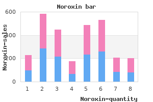
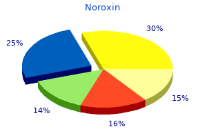
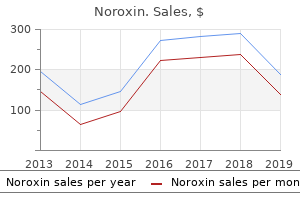
"Order noroxin us, treatment for dogs with food poisoning".
By: Y. Copper, M.S., Ph.D.
Program Director, University of Virginia School of Medicine
It detects the oxygen signals due to the increased blood oxygen levels following increased regional blood flow infection after root canal buy 400 mg noroxin with mastercard. This increase in signal discharge is due to the alteration in paramagnetic properties of deoxygenated hemoglobin antibiotic resistance yahoo generic 400mg noroxin visa. The signals are not specific to the type of the cerebral activity antimicrobial nanomaterials proven 400mg noroxin, which could be excitatory or inhibitory antibiotic 93 7158 buy noroxin 400 mg lowest price. It has the advantage in that it can be repeated, which is of particular use in research studies [30]. The limitations are that the temporal resolution results in a delay of about 10 seconds, and the magnetic sequences deployed make it unsuitable in patients with a cardiac pacemaker or spinal cord stimulator implant. They can be used to measure cerebral blood flow, glucose consumption, or receptor distribution, mostly for opioid receptors in pain conditions. It has limited spatial resolution and the outcome results are provided as complicated data, which require expertise and time for interpretation. All of the imaging techniques involve psychophysical testing; initially scanned under controlled conditions, then imaged following painful stimuli. The cerebral areas showing changes following noxious stimuli are identified by digital subtraction analysis. A commonly used stimulus design involves "thermal grid pain illusion" which has alternating hot and cold bars. When separately activated, the bars are perceived as innocuous sensation (non-painful, either hot or cold) but when activated together, are perceived as noxious. The specific cerebral areas activated give a clue to the areas that may be involved 19 Section 1: the Clinical Presentation of Neuropathic Pain in the pain information process, termed the "pain matrix" [16]. It is important to understand that these areas do not represent "pain centers" as they are also stimulated in other non-painful cognitive states. All areas in the brain are stimulated bilaterally in response to the noxious stimulus except the primary somatosensory cortex (S1), which has a response contralateral to the side of stimulus. The affective and attentional components to pain are thought to be processed in the prefrontal, parietal, and cingulate cortex, whereas the S1, lateral thalamus, and posterior insula are involved with the sensory-discriminative aspect of pain perception. Insular cortex was seen to be closely involved in the autonomic response to pain [15,16]. Although the studies looking into this association are small in number, there is convincing evidence to correlate brain changes with neuropathic pain. A decreased blood flow to the contralateral thalamus was observed with unilateral spontaneous neuropathic pain and reversal of these changes was observed with analgesic treatment. This has led to an assumption that regional hypoperfusion could be used for diagnosis of neuropathic pain and restoration of regional cerebral blood flow as an indicator for treatment efficacy [16]. This has also led to the suggestion that a shift in hemispheric balance might be a cause for allodynia. Studies have also demonstrated the efficacy of psychological intervention in treatment, with distraction and hypnosis therapy producing attenuation of the signals in S1 and cingulate cortex; although not the thalamus [26]. Another interesting observation with the imaging techniques is the demonstration of the ability of the S1 to reorganize the cortical homunculus following painful injuries such as amputation. The S1 area usually involved with afferent input from the amputated limb receives input from adjacent areas of the S1 cortex, called cortical remapping. This is thought to be associated with phantom limb phenomena and pain, as the reversal of these changes and a reduction in pain were seen when a prosthetic limb was utilized. The imaging of opioid receptors, both in central and peripheral neuropathic pain, has demonstrated abnormalities in terms of a decrease in receptor availability and drug binding with pain processing circuitries [33]. In future, better understanding of the effect of analgesics on brain activity could lead to the development of novel therapeutic strategies. Therefore the evidence to support them for diagnostic procedures is low, but they are still being encouraged to be utilized more in neuropathic pain [16]. Autonomic tests Neuropathic pain can be associated with features of autonomic dysfunction such as skin color and temperature changes, sweating abnormalities, and edema. Therefore tests which study skin thermoregulation, sudomotor, and cardiovagal functions have been used to assess autonomic functions [Table 2. Autonomic function testing relies on indirectly accessing the function of unmyelinated postganglionic fibers, which cannot be tested directly by conventional neurophysiological techniques [15,16]. The thermometer probes are attached to the distal part of the affected and contralateral normal limbs. This test has a low specificity and sensitivity as there may be no difference in baseline temperature between the two sides [37,38]. It involves the transdermal administration of 10% acetylcholine by iontophorosis under constant 2 mA current flow for 5 minutes [39]. Sweat production is measured afterwards at four sites; medial distal forearm, proximal lateral leg, medial distal leg, and dorsum of the foot. In painful small fiber neuropathy, if reduced or absent sweat production occurs, it may imply autonomic system involvement in pain generation.
The effects of sleep deprivation on pain inhibition and spontaneous pain in women antibiotics and milk purchase noroxin with american express. Individual variation in rapid eye movement sleep is associated with pain perception in healthy women: preliminary data bacteria and blood in urine buy cheap noroxin. Sequential daily relations of sleep antibiotic ointment packets cheap noroxin amex, pain intensity antibiotics weight loss buy noroxin 400 mg low price, and attention to pain among women with fibromyalgia. Retarded disengagement from pain cues: the effects of pain catastrophizing and pain expectancy. The anticipation of pain modulates spatial attention: evidence for pain-specificity in high-pain catastrophizers. Dimensions of catastrophic thinking associated with pain experience and disability in patients with neuropathic pain conditions. Self-reported sleep quality and quality of life for individuals with chronic pain conditions. Sleep deprivation and activation of morning levels of cellular and genomic markers of inflammation. Cholinomimetics, but not morphine, increase antinociceptive behavior from pontine reticular regions regulating rapid-eye-movement sleep. Sleep in depressed and nondepressed participants with chronic low back pain: electroencephalographic and behaviour findings. Decreased sleep spindles and spindle activity in midlife women with fibromyalgia and pain. Acute intravenous administration of morphine perturbs sleep architecture in healthy pain-free young adults: a preliminary study. Cognitive behavioral therapy for treatment of chronic primary insomnia: a randomized controlled trial. Clinical significance and predictors of treatment response to cognitivebehavior therapy for insomnia secondary to chronic pain. Prevalence of self-reported neuropathic pain and impact on quality of life: a prospective representative survey. Association between cingulum bundle structure and cognitive performance: an observational study in major depression. Immune regulation of central nervous system functions: from sickness responses to pathological pain. Painful diabetic neuropathy is more than pain alone: examining the role of anxiety and depression as mediators and complicators. The coexistence of neuropathic pain, sleep, and psychiatric disorders: a novel treatment approach. The relationship between cytokines and pain/ depression: a review and current status. Systematic review and metaanalysis of randomized controlled trials of cognitive behaviour therapy and behaviour therapy for chronic pain in adults, excluding headache. Assessment and treatment of psychosocial comorbidities in patients with neuropathic pain. Mood and anxiety disorders associated with chronic pain: an examination in a nationally representative sample. Depression-anxiety relationships with chronic physical conditions: results from the World Mental Health Surveys. A randomized, double-blind, placebo-controlled, fixed-dose, multicenter study of pregabalin in patients with generalized anxiety disorder. The Pittsburgh Sleep Quality Index: a new instrument for psychiatric practice and research. An inventory for measuring 332 Chapter 27: Impact of chronic pain upon anxiety, sleep, and mood dimensions depression. The impact upon health-related quality of life in these patients is greater than the sufferers of chronic conditions such as heart failure, cancer, chronic obstructive pulmonary disease, and motor neuron disease. This indicates a significant health burden for patients suffering with neuropathic pain [1] and for the society supporting them. Health-related quality of life Disease itself, as well as the side effects of treatment, affect quality of life. This encompasses physical and mental health along with the other aspects of life that affect health in its broadest sense. Every person has a different perception of what is important to them in life and hence defines quality of life differently. Quality of life is a multidimensional construct that includes biological, psychological and social domains. The overall quality of life depends on various aspects, including employment, housing, neighborhood, cultural, and spiritual values of the Impact of chronic neuropathic pain on quality of life Pain is an individual experience, a subjective feeling that is difficult to measure. An extensive crosssectional survey of Finnish adults revealed chronic pain as an independent predictor of self-rated poor health [6]. Sometimes symptoms are cured, but the side effects of treatment significantly affect QoL. For example interventions for pain relief may produce drowsiness, nausea or memory impairment which may potentially impair physical and emotional function and exacerbate comorbid symptoms, which thereby offset the therapeutic benefit. Pain Physical functioning Emotional functioning Participant ratings of global improvement Symptoms and adverse events Participant disposition Enjoyment of life in general Fatigue Emotional well-being Weakness Staying asleep at night Other aspects of daily life such as travel and getting around in the community.
Meningiomas of the jugular foramen virus children discount noroxin 400mg without prescription, on the other hand treating dogs for dry skin cheap noroxin 400 mg, rarely cause expansion of the jugular foramen and instead tend to infiltrate the bone or occasionally (but not commonly) result in hyperostosis virus bacteria purchase 400 mg noroxin visa. Of note virus mega brutal noroxin 400mg without a prescription, the margins of the nonexpanded jugular tend to appear irregular because of small areas of cortical loss. When meningiomas primarily arise in the jugular foramen, the extraaxial component of the lesion tends to demonstrate an en-plaque pattern of growth, and the pattern of underlying bony infiltration tends to be extensive. When bony invasion is evident, an enhancing permeative-sclerotic pattern may be seen. Internal or peripheral calcification of the tumor, when present, often can be a helpful key to diagnosis. Although meningiomas generally are avidly enhancing lesions, the absence of internal flow voids are another helpful feature differentiating them from jugular foramen paragangliomas. When the diagnosis of jugular foramen meningioma is being considered, attention to the dura may reveal important clues to this diagnosis, such as the identification of dural tails or the already mentioned en-plaque dural growth. The diagnosis of jugular foramen metastasis can be entertained with a greater degree of suspicion when indicated by a history of a primary malignancy and/or the identification of multiple lesions. Metastatic involvement of the jugular foramen is most often associated with an aggressive permeative/destructive pattern of bone involvement. The appearance of the enlarged nerve indicates a lesion intrinsic to the optic nerve, such as an optic nerve glioma. A broad-based, homogeneously enhancing mass is noted in the left posterior fossa, consistent with an additional meningioma. In adult populations, the usual presenting symptom is rapidly progressive vision loss. When diagnosed in an adult, the lesion usually is a malignant astrocytoma or glioblastoma and carries a poor prognosis. The classic imaging finding is tubular, noncalcified enlargement of the optic nerve. The classic imaging finding is the tram track sign, associated with linear calcifications or enhancement parallel to the optic nerve. Fat-suppressed magnetic resonance imaging is preferred to identify the lesion and determine the extent of disease. In addition to optic nerve gliomas, differential considerations include optic neuritis, orbital sarcoid, and idiopathic orbital inflammatory disease, and thus the clinical history may be helpful. Both types of lesions may result in adjacent osseous scalloping or expansion of the bony canals. Differential considerations include sarcoidosis, demyelinating optic neuritis or perineuritis, orbital inflammatory disease of the optic nerve, orbital schwannoma, lymphoma, hemangiopericytoma, and optic nerve metastasis. Edema of the optic disc often is seen initially with meningiomas, and the disc subsequently appears to be atrophic. Each cavernous sinus communicates with the contralateral side, the pterygoid plexus, the inferior petrosal sinus, and the superior ophthalmic veins. Asymmetric enhancement of the cavernous sinus can be the result of thrombosis, a mass, inflammatory processes such as a sarcoid, Tolosa-Hunt syndrome, or a fungal infection. An important imaging finding may be enlargement of a superior ophthalmic vein as a result of either a fistula or thrombosis. Predisposing factors to spontaneous rupture include fibromuscular dysplasia, Ehlers-Danlos syndrome, and pseudoxanthoma elasticum. Associated venous hypertension can result in ocular signs and symptoms of proptosis, chemosis, conjunctival injection, cranial nerve palsies, and visual deficits. Cerebral complications include intracranial hemorrhage, increased intracranial pressure, and vascular steal. Imaging demonstrates enlargement of the extraocular muscles, the superior (and/or inferior) ophthalmic vein, and the ipsilateral cavernous sinus. Digital subtraction angiography is essential to confirm the diagnosis and classify the fistula and to identify sites of venous drainage. Other common symptoms include orbital pain, periorbital edema, chemosis, ptosis, ophthalmoplegia, and visual loss. In persons with septic thrombosis, systemic signs of sepsis occur as a late finding. Because the veins communicating with the cavernous sinus do not have valves, bacteria from facial, paranasal sinus, and odontogenic infections can pass into the cavernous sinus Dilated Superior Ophthalmic Vein/Asymmetric Cavernous Sinus Enhancement 345 A Cor T1 Post B Ax T1 Post C Ax T2 Figure 56-1 A 40-year-old woman with intermittent trigeminal neuralgia. Coronal T1 postcontrast images, axial T1 postcontrast images, and axial T2-weighted images demonstrate a homogeneously enhancing, dural-based, extraaxial mass in the left cavernous sinus consistent with cavernous sinus meningioma. Imaging findings include enlargement of the cavernous sinus with filling defects and enlargement and thrombosis of the superior and inferior ophthalmic vein. The lacrimal gland is divided into a deeper orbital lobe and a more superficial palpebral lobe that is one third to half the size of the orbital lobe (Figure 57-1). In general, neoplasms, particularly epithelial neoplasms, have a propensity to involve a single lobe of the lacrimal gland, whereas inflammatory, autoimmune, and lymphoproliferative processes tend to involve the entire gland. Lacrimal lesions first should be evaluated for focal or diffuse involvement and can be divided into epithelial and nonepithelial lesions. Epithelial lesions, which make up 40% to 50% of lacrimal gland lesions, tend to involve the deeper orbital lobe and therefore have a greater propensity to grow posteriorly. Malignant epithelial neoplasms have histologies similar to those seen in the salivary glands. Nonepithelial lesions often present with involvement of both lobes of the lacrimal gland and can be generalized into those typically having unilateral versus bilateral involvement. Acute dacryoadenitis often is related to a bacterial or viral infectious process commonly presenting in children and young adults with erythema, tenderness, and discharge.
Syndromes
Abnormal EKG
Bleeding prostate
Hypothyroidism
Venipuncture
Pollens
Ask your doctor which drugs you should still take on the day of your surgery.
Smith-Lemli-Opitz syndrome
The cyst also may be mobile within the ventricular system and migrate between examinations yeast infection 9dpo purchase noroxin 400 mg fast delivery. Their location in the anterior third ventricle predisposes them to obstruction of the foramina of Monro and acute hydrocephalus (which may be intermittent) antibiotics for dogs amoxicillin dosage buy noroxin paypal, with rare cases of sudden death reported antibiotics for strep viridans uti buy noroxin 400mg on line. They usually densely enhance and frequently are marked by cysts oral antibiotics for sinus infection order noroxin in united states online, calcification, and hemorrhage. Other Differential Diagnoses Discovered in 1998 and named for its microscopic resemblance to chordomas, all chordoid glioma lesions described to date have been located in the third ventricle. Choroid Plexus Papilloma and Carcinoma Masses that arise from the suprasellar and pineal regions often appear to be arising from the third ventricle. Craniopharyngiomas and germinomas should be considered in the differential diagnosis when an anterior third ventricular mass is visualized. Pineal region tumors and germinomas should be considered when a posterior third ventricular mass is visualized. Restricted diffusion (low apparent diffusion coefficient) is typical because of high tumor cellularity. Extension through the foramina of Magendie (~60%) and Luschka (~15%) frequently occurs. In children, medulloblastomas and ependymomas are the most common masses found in the fourth ventricle. Other pediatric posterior fossa primary neoplasms, including pilocytic astrocytoma and brainstem glioma, occasionally may grow exophytically into the fourth ventricle and mimic a mass of ventricular origin. Although a hemangioblastoma rarely arises within the fourth ventricle, it should be considered in the setting of von Hippel Lindau disease. Subependymomas are often small (<2 cm), but mean lesion size is approximately 4 cm in symptomatic patients with hydrocephalus. Choroid Plexus Papilloma and Carcinoma Choroid plexus tumors occur in the lateral ventricle (50% of cases), fourth ventricle (40% of cases), and third ventricle (10% of cases). Choroid plexus tumors occur most commonly in young adults (median age, 23 years) when they appear in the fourth ventricle. In adults, a cerebellar hemispheric location is more common; this finding is thought to be related to the superolateral migration of undifferentiated cells with oncogenic potential originating in the posterior medullary velum. In less clear-cut cases, a careful search for parenchymal or cisternal disease will increase diagnostic specificity. Ependymomas are differentiated from medulloblastomas by a higher likelihood of demonstrating calcifications, hemorrhage (T2 imaging), higher diffusivity, and "plastic" tumor behavior with extrusion through the foramina of Magendie and Luschka. Thought to arise from subependymal glia, subependymomas are benign (World Health Organization grade I), often incidental tumors that are most commonly seen in middle-aged or elderly men. Approximately 10% demonstrate an admixture of ependymoma on histopathologic examination. Modern microsurgical techniques yield good outcomes, even with subtotal resection. A sagittal postcontrast fat-saturated image demonstrates an irregular, mildly thickened, and mildly nodular rim of enhancement. Although solid or vascular lesions related to the optic nerves or chiasm, the circle of Willis, the hypothalamus, or pituitary infundibulum present at this site, the identification of a cystic-appearing lesion significantly narrows the differential diagnosis. When evaluating a cystic suprasellar lesion, the presence of nodular or frankly solid enhancing components suggests a neoplastic lesion, the most common of which is an adamantinomatous craniopharyngioma. Craniopharyngiomas may be sellar and suprasellar, exclusively suprasellar, or purely intrasellar. They demonstrate a bimodal distribution, with two thirds of them presenting in childhood/adolescence and a second smaller peak generally presenting in middle to late adulthood. Childhood craniopharyngiomas tend to be adamantinomatous and present as predominantly cystic or solid and cystic lobulated masses with hemorrhage and calcification. Adult craniopharyngiomas tend to be papillary and present more often as predominantly solid or mixed solid and cystic masses. As noted, careful inspection for the presence of nodular or frankly solid enhancing components should be performed. Nodular or rimlike calcification involving cyst walls or solid components is highly suggestive of this diagnosis. The hallmark characteristics of predominantly cystic craniopharyngiomas are calcification, cyst formation, and nodular and/or rim enhancement. Furthermore, other less common neoplastic lesions that typically present as cystic and solid masses, such as hypothalamic/optic pathway juvenile pilocytic astrocytomas, should be considered in the appropriate clinical setting. Approximately 80% of these lesions are seen between the anterior and posterior pituitary lobes. On imaging, they typically appear as smoothly contoured, spherical or ovoid, nonenhancing lesions, although a thin rim of peripheral enhancement occasionally can be present. However, 102 Brain and Coverings solid or nodular enhancement such as that seen in craniopharyngiomas should not be present. Despite the variable signal intensity of different lesions, the signal intensity is generally homogeneous. Importantly, in up to 40% of lesions the homogenous signal intensity is disrupted by the presence of an intracystic T2 hypointense nodule (protein and cellular debris), the identification of which is particularly suggestive of this diagnosis. Although they occur anywhere along the neuraxis, 10% to 15% present in the suprasellar region. The morphology of these lesions provides further distinguishing features from arachnoid cysts. Epidermoids generally exhibit lobulated, crenulated, and irregular margins compared with the smooth walls of arachnoid cysts.
Buy discount noroxin 400 mg on-line. Towel Can Do Miracles For Neck Pain - Dr Mandell.